
AFM – Delivering Profound Insights in Microbiology
Wednesday, December 3 | 8AM PST | 11AM EST | 5PM CET
We warmly invite you to join us and our special guest speaker Dr. Laia Pasquina Lemonche of the University of Sheffield, UK, for this webinar on “AFM – Delivering Profound Insights in Microbiology”.
Atomic force microscopy (AFM) is an advanced multiparametric imaging technique. It delivers comprehensive insights into the nanomechanical properties of biomolecules, cells, bacteria, and viruses, enabling the characterization of their structure and morphology and the quantification of, for example, pathogen-host cell interactions and immune response.
AFM can be seamlessly combined with advanced optical microscopy techniques, like confocal and super-resolution microscopy, for enhanced imaging capabilities and profound insights into molecular processes.
Bruker BioAFMs have been optimized for use in microbiology and virus research, enabling direct access to pathogens and infectious agents in BSL facilities.
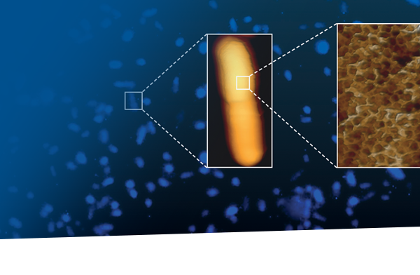
In her talk, “Reaching Unprecedented Imaging Resolution of Bacterial Cell Wall with AFM” Dr. Pasquina Lemonche will speak on using AFM to decipher the 3D architecture of the polymer peptidoglycan found in bacterial cell walls.
The webinar will include a demonstration LIVE from our labs in Berlin, illustrating the capabilities of Bruker’s NanoWizard BioAFM platform.
Special Guest Speaker

Dr. Laia Pasquina Lemonche
The University of Sheffield, UK
Dr. Laia Pasquina Lemonche is an Early-Career-Award Wellcome Trust Fellow. She has a degree in Physics and received a Master’s in Nanobiotechnology from the Autonomous University of Barcelona (UAB) in 2016. In 2020, she obtained a PhD in Biophysics from the University of Sheffield where she focused on deciphering the molecular architecture of gram-positive bacterial cell walls using AFM. She went on to become a Research Associate in Professor Jamie Hobbs’ group until 2024, pushing the limits of resolution of biological material. Currently, her group focuses on using correlative AFM and STORM to study Streptococcus pneumoniae peptidoglycan and developing novel software approaches for microscopy image analysis.
Talk Title & Abstract
Reaching Unprecedented Imaging Resolution of Bacterial Cell Wall with AFM
The cell wall of bacteria is mainly composed of a disordered polymer called peptidoglycan. As this is a non-crystalline material, its three dimensional architecture cannot be determined with the traditional techniques often used in microbiology, such as cryo-EM or NMR. For this reason, the Hobbs and Pasquina groups at the University of Sheffield have, for many years, been using AFM to decipher the three-dimensional architecture of this un-ordered material. Using Bruker’s atomic force microscopes, they have been able to push the imaging resolution of this highly deformable biological material to unprecedented levels. In her talk Dr. Pasquina Lemonche will share AFM methods and present landmark studies.


